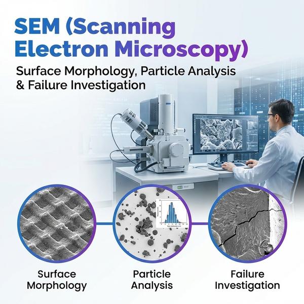
At Dextrose Technologies Pvt. Ltd., we provide SEM (Scanning Electron Microscopy) for high-resolution imaging and detailed surface characterization of materials and biological samples (as applicable). SEM helps you visualize surface morphology, topography, particles, and defects with clarity—supporting R&D, quality investigations, and failure analysis. Surface morphology and topographical analysis We capture high-magnification images to evaluate surface texture, porosity, cracks, coatings, and microstructural features. SEM is ideal for comparing batches, processing conditions, and material finishes. Particle shape and size evaluation We image particles to assess shape, agglomeration, and size distribution trends (image-based), supporting powders, pigments, fillers, nanomaterials (where applicable), and formulation studies. Failure and defect analysis We support root-cause investigations for defects such as fractures, delamination, inclusions, corrosion features, and surface contamination—providing image evidence and interpretation notes to guide corrective actions. Deliverables * High-resolution SEM images (multiple magnifications) * Observation summary highlighting key morphological features * Optional: EDX elemental mapping/spot analysis (if required) * Report-ready documentation for R&D or QC investigations Get started Share your sample type, objective (morphology/particles/failure), and preferred magnification range. We’ll recommend the best SEM imaging plan and deliverables.

Hyderabad
08048047718
+919686928701
 Chat with us